
Anatomy 101
With the start of school, it seemed an appropriate time to review anatomy. Based on frequent questions, this article briefly covers some of the points of anatomy that would serve for a deeper understanding in a Pilates practice and beyond. Going from the inside out, we’ll look at bones, ligaments, muscles, tendons and fascia.
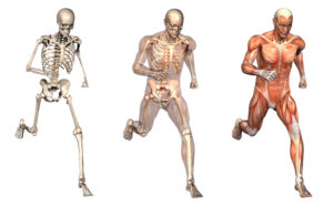
Bones:
At the deepest level, the bones are the framework for the body. Bones protect important areas such as the brain, spinal cord and heart. Bones provide structure for mobility by giving the muscles something they can attach to and leverage in order to move.
Because bones are such a strong structure, it’s easy to think of them in a stiff way. It’s remarkable to consider the human frame is actually living tissue! At the marrow of the bone, red and white blood cells are manufactured. The bone itself either grows denser or more porous as the bone cells lay down bone or reabsorb bone. This is an important process. In the next newsletter, we’ll examine bone health more closely, specifically looking at osteoporosis and how Pilates can help maintain strong bones.
Ligaments:
The bones wouldn’t make as effective a frame without the ligaments. Ligaments connect the bones together. This forms the joints. Some ligaments allow certain movements and other ligaments might prevent movement in a certain direction altogether.
Muscles:
Muscles move the body. Internally, there are cardiac and smooth muscles (lining the esophagus, intestines,…) which are “involuntary”. More superficially are skeletal muscles which are “voluntary muscles” or under conscious control. When a skeletal muscle contracts, it brings the bones closer together. For example, when the biceps contracts it moves the upper and lower arm bones towards each other.
The two categories of skeletal muscles are fast and slow twitch muscle fibers (light and dark meat). Fast twitch powers explosive, quick movements but these muscle fibers fatigue swiftly. Slow twitch have more endurance but not as much force.
Muscles break down into smaller and smaller bundles. The outermost layer called the epimysium is what most people think of as the muscle and at each end are tendons that attach the muscle to the bone. The perimysium is the next layer and consists of bundles of fibers. The smallest layer is the endomysium and contains myocytes or the single muscle fiber. At the cellular level muscles have protein filaments, actin and myosin, that slide past each other causing the actual muscular contraction.
Tendons:
Each layer of the muscle (epimysium, perimysium and endomysium) contributes fibers that create the muscle tendon. Tendons connect muscle to bone. Tendons are the beginning (origin) and end (insertion) of a muscle. In the middle is the “belly of the muscle”, where the muscular contraction occurs.
Fascia:
The layers of muscles are each covered in fascia. While muscles can be dissected and considered to neatly begin and end, the fascia is continuous throughout the body. Fascia covers not only muscle but also internal organs.
Conclusion:
This “bare bones” article on anatomy briefly touched on bones, ligaments, muscles, tendons and fascia. More in depth coverage could be given to each of these topics, not to mention subjects like physiology, organs and the brain/body connection. This focused article intended to give simple, clear body basics, Anatomy 101!